The challenge to distinguish between patients with hereditary RBC disease from iron deficiency and other diseases
Although haematology analysers are not able to directly determine morphologic abnormalities, the biology of RBC diseases is reflected in the red blood cell indices and in advanced red blood cell parameters. Intelligent interpretation of blood counts and algorithms taking advantage of disease features present a fast and sensitive solution to identifying certain RBC diseases, with the ability to distinguish them from other aetiologies that result in abnormal red blood cell indices. In a series of two studies, Nivaggioni et al. were able to provide a holistic approach and described a reliable algorithm that distinguished several hereditary red blood cell diseases (including heterozygous haemoglobino-pathies, sickle cell disease, hereditary spherocytosis and Southeast Asian ovalocytosis) from patients with IDA (an acquired condition) or other conditions.
Pharmacist and medical biologist Dr Vanessa Nivaggioni manages the haematology laboratory at University Hospital de la Timone in Marseille, France.
The hospital is a reference centre for major sickle cell syndromes, thalassaemias and other rare diseases of the red blood cells and erythropoiesis. The hospital provides care for both adult and paediatric patients.

Firstly, thanks a lot for your time and congratulations on your publications. In your initial publication, you described a new CART tree* for predicting hereditary RBC diseases using various parameters from the XN analyser to guide further investigation and potential treatment outcomes.
Can you describe the various conditions the decision tree can predict and why these conditions present a challenge in the laboratory?
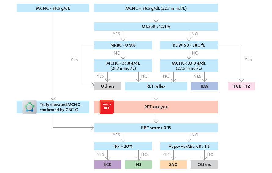
This algorithm can correctly differentiate patients with hereditary RBC diseases such as heterozygous haemoglobinopathy (HGB HTZ: thalassaemia, heterozygous sickle cells), sickle cell disease (SCD: homozygous and combined heterozygous sickle cell disease with thalassaemia trait) and hereditary spherocytosis (HS) from iron deficiency anaemia (IDA) and other patients. IDA patients must be correctly identified so as not to interfere in the detection of RBC disease. The aim is to guide the use of complementary examinations for the diagnosis: blood smear, iron status, haemoglobin electrophoresis and eosin-5-maleimide binding test (EMA test).
Red blood cell diseases, in particular haemoglobinopathies, are frequent and widespread worldwide. Heterozygous subjects are generally asymptomatic but homozygous patients are exposed to severe and even fatal complications. Prevention of homozygous haemoglobinopathies is achieved by screening for asymptomatic carriers. Our algorithm was created with this in mind. As such it addresses all kinds of laboratories.
What are the applications and parameters used on the XN analyser that enabled the creation of this decision tree?
As previously mentioned, parameters and their cut-off values were selected by the CART method for this two-step decision tree. The statistical robustness of MicroR is favourable and used to directly distinguish between IDA, RBC disease and other causes of RBC abnormalities instead of MCV and MCH, two traditional parameters used for anaemia classification. Because of the heterogeneity of our population, MicroR was not sufficient alone to predict an outcome. Other parameters are also required. RDW-SD measures absolute anisocytosis and helps to differentiate thalassaemia trait (without anisocytosis) from IDA and SCD with MicroR > 12.9%. NRBC% is typical of SCD and MCHC helps for HS, SCD (elevated MCHC), IDA and HGB HTZ (low MCHC). Adding a RET channel measurement is requested in some cases to properly discriminate some RBC diseases from other patients. In fact, SCD and HS patients have a positive RBC Score due to increased reticulocytes count with or without FRC% and an elevated (SCD) or low (HS) IRF% in contrast with other patients (negative RBC score).
Are you now using the RBC decision tree in your clinical routine? If so, how is it perceived by clinicians?
Right now, clinicians don't see it because they were already used to having internal criteria for adding haemoglobin electrophoresis but, using the RBC tree, these additions are now much better targeted and offer better sensitivity and specificity.
A major concern we have is that we are missing the addition of iron status when there is a suspected IDA. The reason is that the haematology laboratory is not connected to the chemistry one for the iron assessment, but we are working on this change.
Can you give us an overview of the further developments that you’ve been working on since the initial publication?
Despite the good RBC disease classification rate of this decision tree, patients presenting with Southeast Asian ovalocytosis (SAO) were misclassified as patients with no RBC disease. Indeed, these patients show in general an increased MCHC overlapping 365 g/L and simultaneously a severe difference in haemoglobin whereby HGB-O appears much lower than photometric haemoglobin. As a consequence, via the CBC-O application, these samples require a haemoglobin interference check with a clear plasma. This can lead to confusion amongst users in deciding which haemoglobin value to correctly report. A second study has been achieved to complete this established decision tree to improve the detection of SAO patients. The HYPO-He/MicroR ratio correctly identifies 100% of SAO cases in the presence of an increased MCHC.
How has implementing this decision tree in the Sysmex Extended IPU solution benefitted your laboratory in terms of clinical value and workflow efficiency?
The decision tree rules are all integrated into the Sysmex Extended IPU solution. This means that the rules are automatically triggered upon receiving the first blood count results of an unknown patient or with an old blood count (older than 30 or 90 days depending on the suspected pathology). In some cases, the addition of the RET channel and/or a blood smear is necessary and done automatically. This saves the operator a lot of time. Moreover, the generated blood smear is targeted which helps the cytologist for microscopy review. In our routine, the smears are focused on SCD and HS suspicion as a preliminary control while heterozygous haemoglobinopathies are sent directly for electrophoresis. This also offers a better focus on RBC smear management.
Dr Nivaggioni we would like to thank you very much for taking the time for this interview.
References
- Berda-Haddad Y et al. (2017): Increased mean corpuscular haemoglobin concentration: artefact or pathological condition? Int J Lab Hematol. 39(1):32-41
- Nivaggioni V et al. (2020): Use of Sysmex XN-10 red blood cell parameters for screening of hereditary red blood cell diseases and iron deficiency anaemia. Int J Lab Hematol. 42(6):697-704. 2.
- Nivaggioni V et al. (2021): Detection of Southern Asian Ovalocytosis with Sysmex XN-10: A complement to the decision tree previously described. Int J Lab Hematol. Online ahead of print
* The published two-step decision tree described in this interview can be implemented as a ruleset in the Sysmex Extended IPU and is promoted under the term ‘RBC Disease Manager’ by Sysmex. The implementation of the RBC Disease Manager in Extended IPU achieves complete automation of the workflow for handling patient samples exhibiting the RBC diseases outlined in this interview.