Why digitise pathology?
Demographic and economic developments and fast-paced progress in human medical research mean that histopathological practice must adapt to meet new requirements. Due to shifting demographics, the number of samples laboratories receive continues to rise. In addition, more diagnostic markers and the increasing complexity of cases result in increased staining per case. Falling rates of board certification pose a significant challenge; employment opportunities therefore need to become more flexible, both in terms of working hours and location.
By working together, we will find the ideal solution to make your laboratory fit for the future.
Digitization provides fast and flexible access to all data
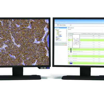
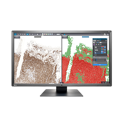
Keep everything in view with the digital cockpit
- All information on a specific case file is displayed at all times in the pathologist’s cockpit
- The site-independent tool enables pathologists to access digital slides from any location – and make diagnoses from their home office
- The database of digitized slides and the virtual microscope can be seamlessly integrated in an existing LIS
- Digitization standardises and optimises the workflow, saving valuable time and providing safety
Site-independent tumour conferences modernise communication
Overcoming geographic separation
- The ability to analyse digitized tissue sections collaboratively from different locations saves time and reduces costs
- High-quality digital tissue sections with diagnostic information can be presented as part of a tumour conference
- Macro shots, archive images, special staining and quantification results can be easily shared, facilitating a rapid exchange between experts
High quality management standards provide safety
Support for accreditation processes
- The digital workflow simplifies the process descriptions in the QMS manual
- Large quantities of data and images are automatically stored in the right place and can be traced at any time
- In accordance with legal provisions, case files can be archived in an automated, standardised manner
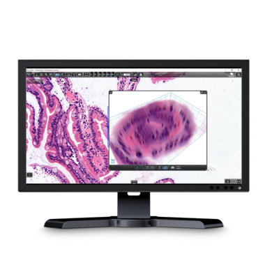
Digital slides expand diagnostic possibilities
Making the diagnostic process easier
- Digital tools, such as annotation, marking and comment functions, simplify morphological diagnosis
- Various tissue staining images and macro shots can be displayed and analysed in parallel
- The digital archive provides fast access to all earlier slides in a given case
- Digital quantification of diagnostic markers supports the diagnostic process and establishes standardised criteria for analysis